Abdominal Anatomy ~ Medically Accurate Illustration Of The Abdominal Anatomy Stock Photo Alamy. For the sake of brevity, the various organs will be not discussed in detail. Abdomen, in human anatomy, the body cavity lying between the chest or thorax above and the pelvis below and from the spine in the back to the wall of abdominal muscles in the front. We'll identify as many organs as we can, see how they fit into the. Abdominal wall anatomy that is clinically pertinent to the surgeon, focusing primarily on the structures of the anterior abdominal wall, will be reviewed. The abdominal aorta enters the abdomen through the diaphragm at the level of the twelfth thoracic vertebre and continues to just below the umbilical area, where it splits into the right and left common iliac arteries.
Then liver & spleen) palpate 4 quadrants abdomen (superficial then deep) assess for kidney area pain (cvat) wash hands time target: It also contains the spleen. The regions occupied by stomach are epigastric, umbilical and hypochondriac regions. Together, these three turn nutrients into usable energy, as well as help dispose of solid waste. Abdomen, in human anatomy, the body cavity lying between the chest or thorax above and the pelvis below and from the spine in the back to the wall of abdominal muscles in the front.
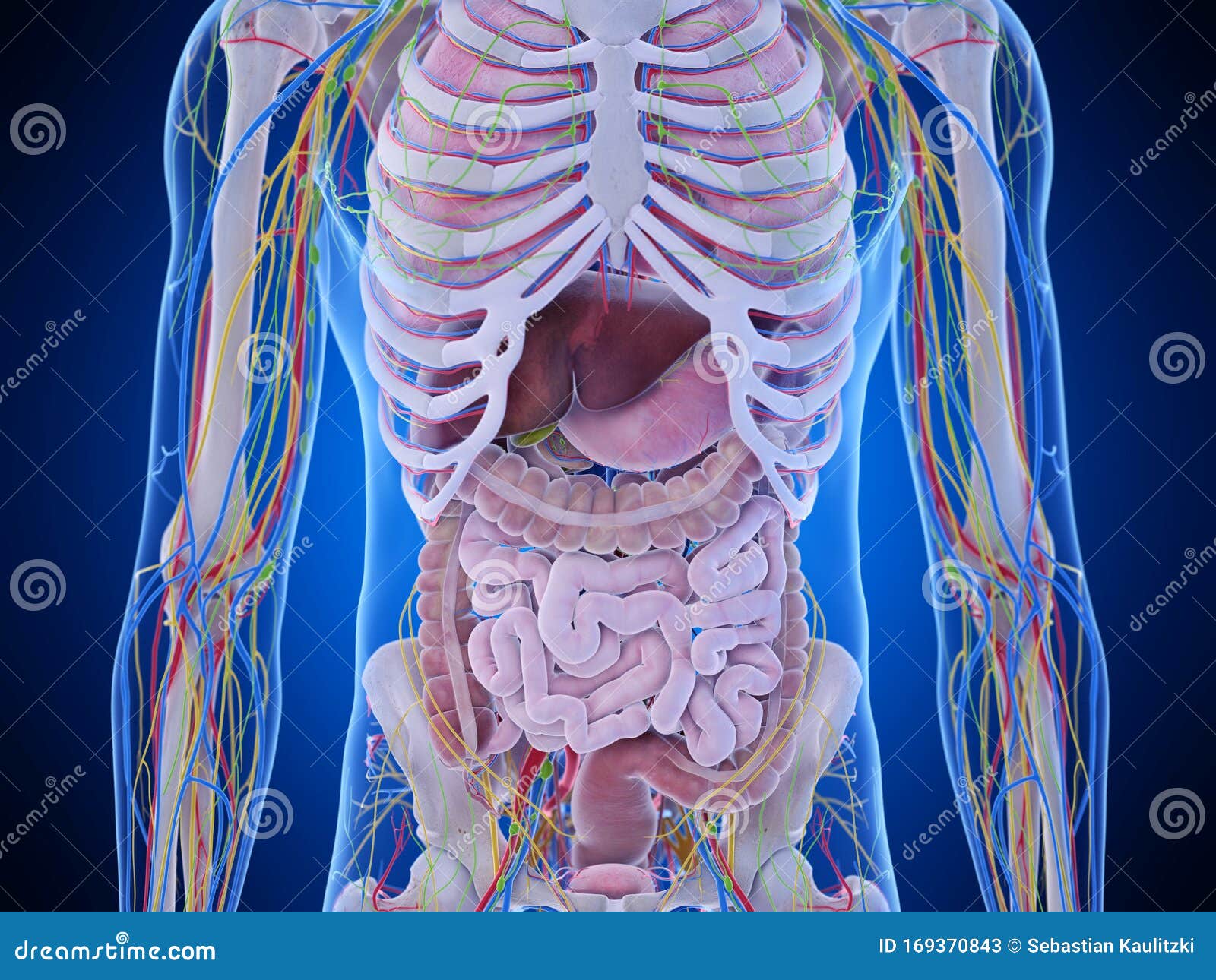
The Abdominal Anatomy Stock Illustration Illustration Of Accurate 169370843 from thumbs.dreamstime.com The region occupied by the abdomen is called the abdominal cavity, and is enclosed by the abdominal muscles at front and to the sides, and by part of the vertebral column at the back. The majority of these organs are encased in a protective membrane termed the peritoneum. The transverse abdominal muscle wraps around the torso from front to back and from the ribs to the pelvis. It is an artery, meaning that it carries blood away from the heart. Inferiorly the abdomen is open to the pelvis, communicating through the superior pelvic aperture (pelvic inlet). Together, these three turn nutrients into usable energy, as well as help dispose of solid waste. The abdomen is the body region found between the thorax and the pelvis. The component of the urinary system, kidney and the ureter.
The abdomen is the front part of the abdominal segment of the trunk.
These organs are held together loosely by connecting tissues. Abdominal anatomy muscles 12 photos of the abdominal anatomy muscles abdominal muscles anatomy and function, abdominal muscles anatomy diagram, abdominal muscles cross sectional anatomy, deep abdominal muscles anatomy, lateral abdominal muscles anatomy, human anatomy, abdominal muscles anatomy and function, abdominal muscles anatomy diagram. The normal anatomy or organs imaged in a standard abdominal examination is explained below. Together, these three turn nutrients into usable energy, as well as help dispose of solid waste. These two apertures, together with abdominal walls, bound the abdominal cavity. The abdomen (colloquially called the belly, tummy, midriff or stomach) is the part of the body between the thorax (chest) and pelvis, in humans and in other vertebrates. Observation, auscultation, percussion, and palpation. We're going to take apart a plastic anatomy model and see what we can find in the abdomen. If you plan to enter a healthcare profession such as nursing, this is something you'll use on the job when performing abdominal assessments (and while documenting). The muscle fibers of the transversus abdominis run horizontally, similar to a corset or a weight belt. The region occupied by the abdomen is called the abdominal cavity, and is enclosed by the abdominal muscles at front and to the sides, and by part of the vertebral column at the back. The anterolateral abdominal wallformed of 4 layer skin, fascia, muscles, and peritoneum. The transverse abdominal muscle wraps around the torso from front to back and from the ribs to the pelvis.
In anatomy and physiology, you'll learn how to divide the abdomen into nine different regions and four different quadrants. Observation, auscultation, percussion, and palpation. For the sake of brevity, the various organs will be not discussed in detail. The abdomen is the part of the body that contains all of the structures between the thorax (chest) and the pelvis, and is separated from the thorax via the diaphragm. The majority of these organs are encased in a protective membrane termed the peritoneum.
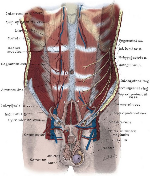
Anatomy Of The Lower Urinary Tract And Male Genitalia Abdominal Key from abdominalkey.com The regions occupied by stomach are epigastric, umbilical and hypochondriac regions. The component of the urinary system, kidney and the ureter. The major components of the abdominal exam include: Abdominal wall anatomy that is clinically pertinent to the surgeon, focusing primarily on the structures of the anterior abdominal wall, will be reviewed. The major organs of the abdomen include the small intestine, large intestine, and stomach. The abdominal aorta enters the abdomen through the diaphragm at the level of the twelfth thoracic vertebre and continues to just below the umbilical area, where it splits into the right and left common iliac arteries. The major organs of the abdomen include the small intestine, large intestine, and stomach. Skin, superficial fascia, muscles and associated fascia, and parietal peritoneum.
The major organs of the abdomen include the small intestine, large intestine, and stomach.
It is bounded superiorly by the xiphoid process and costal margins, posteriorly by the vertebral column and inferiorly by the pelvic bones and inguinal ligament. This muscle doesn't help move the spine or the pelvis, but it does help with respiration and breathing. The abdominal aorta enters the abdomen through the diaphragm at the level of the twelfth thoracic vertebre and continues to just below the umbilical area, where it splits into the right and left common iliac arteries. Abdominal anatomy muscles 12 photos of the abdominal anatomy muscles abdominal muscles anatomy and function, abdominal muscles anatomy diagram, abdominal muscles cross sectional anatomy, deep abdominal muscles anatomy, lateral abdominal muscles anatomy, human anatomy, abdominal muscles anatomy and function, abdominal muscles anatomy diagram. Then liver & spleen) palpate 4 quadrants abdomen (superficial then deep) assess for kidney area pain (cvat) wash hands time target: Abdomen, in human anatomy, the body cavity lying between the chest or thorax above and the pelvis below and from the spine in the back to the wall of abdominal muscles in the front. Common incisions and closure techniques, and prevention and management of wound complications, are discussed elsewhere. In anatomy and physiology, you'll learn how to divide the abdomen into nine different regions and four different quadrants. The abdomen is the part of the body that contains all of the structures between the thorax (chest) and the pelvis, and is separated from the thorax via the diaphragm. We'll identify as many organs as we can, see how they fit into the. Abdominal wall anatomy that is clinically pertinent to the surgeon, focusing primarily on the structures of the anterior abdominal wall, will be reviewed. The anterolateral abdominal wallformed of 4 layer skin, fascia, muscles, and peritoneum. The area occupied by the abdomen is called the abdominal cavity.
The diaphragm marks the top of the abdomen and the horizontal line at the level of the top of the pelvis marks the bottom. The major organs of the abdomen include the small intestine, large intestine, and stomach. The regions occupied by stomach are epigastric, umbilical and hypochondriac regions. These two apertures, together with abdominal walls, bound the abdominal cavity. The abdomen is the body region found between the thorax and the pelvis.
Abdominal Anatomy Injuries Diagram Quizlet from o.quizlet.com Skin, superficial fascia, muscles and associated fascia, and parietal peritoneum. The transverse abdominal muscle wraps around the torso from front to back and from the ribs to the pelvis. The abdominal aorta enters the abdomen through the diaphragm at the level of the twelfth thoracic vertebre and continues to just below the umbilical area, where it splits into the right and left common iliac arteries. Abdomen anatomy the abdomen is comprised primarily of the digestive tract and other accessory organs which assist in digestion, the urinary system, spleen, and the abdominal muscles (shown below). We'll identify as many organs as we can, see how they fit into the. The regions occupied by stomach are epigastric, umbilical and hypochondriac regions. These two apertures, together with abdominal walls, bound the abdominal cavity. It is bounded superiorly by the xiphoid process and costal margins, posteriorly by the vertebral column and inferiorly by the pelvic bones and inguinal ligament.
The major organs of the abdomen include the small intestine, large intestine, and stomach.
For the sake of brevity, the various organs will be not discussed in detail. Auscultation before percussion) and carry different degrees of importance. Together, these three turn nutrients into usable energy, as well as help dispose of solid waste. We'll identify as many organs as we can, see how they fit into the. These organs are held together loosely by connecting tissues. This muscle doesn't help move the spine or the pelvis, but it does help with respiration and breathing. The abdominal wall surrounds the abdominal cavity, providing it with flexible coverage and protecting the internal organs from damage. The muscle fibers of the transversus abdominis run horizontally, similar to a corset or a weight belt. Abdominal wall anatomy that is clinically pertinent to the surgeon, focusing primarily on the structures of the anterior abdominal wall, will be reviewed. If you plan to enter a healthcare profession such as nursing, this is something you'll use on the job when performing abdominal assessments (and while documenting). The major organs of the abdomen include the small intestine, large intestine, and stomach. The region occupied by the abdomen is called the abdominal cavity, and is enclosed by the abdominal muscles at front and to the sides, and by part of the vertebral column at the back. Then liver & spleen) palpate 4 quadrants abdomen (superficial then deep) assess for kidney area pain (cvat) wash hands time target: